Article Text

Statistics from Altmetric.com
One of the important clinical features of asthma is the exercise intolerance due to an exacerbation.1 Yet, to our knowledge, this end point has never been assessed in animal models of asthma. In a mouse model of chemical induced asthma we found early and late alterations in ventilatory function using unrestrained whole body plethysmography.2–,4 Partly because this technique has been heavily criticised,5 we sought to provide another measure of functional impairment in asthma—namely, exercise endurance. This was done by a simple exercise test in which mice were forced to swim against a limited downwards current;6 if they stopped swimming the current pulled them under the water. Swimming endurance time is measured as a proxy of exercise tolerance. Task failure is defined as a period of 5 seconds under water.6
The protocol for sensitising and challenging mice was similar to that used previously,2–,4 with small modifications. Male BALB/c mice (±20 g, 6 weeks old) received dermal applications of 20 µl vehicle (2:3 acetone:olive oil) or 0.3% toluene-2,4-diisocyanate (TDI) on each ear on days 1 and 8. On day 15 they received an intranasal instillation of 10 µl vehicle or 0.1% TDI in each nostril. Treatment with TDI is indicated as 1, while treatment with vehicle is indicated as 0. Thus, the 1/1/1 group consists of mice that received dermal applications of TDI (days 1 and 8) and an intranasal instillation of TDI (day 15), while the 0/0/0 control group consists of mice that received the vehicle on all occasions. All mice had a 5 minute swimming training session on day 13. All swim tests were done in water at 34°C.
On day 15 resting ventilatory function (enhanced pause (Penh)) of each mouse was recorded by whole body plethysmography (EMKA Technologies, Paris, France) for 5 minutes before and 40 minutes after the intranasal instillation. One hour after instillation the mice were made to swim in groups of 2–3 until exhaustion, as defined above. On day 16 (that is, 22 hours after intranasal instillation) the mice had a second swim test. Two hours later methacholine reactivity was assessed by whole body plethysmography. The measurement of the swimming time was not conducted in a blinded manner, but we are confident that this did not influence the results. All experimental procedures were approved by the local Ethical Committee for Animal Experiments.
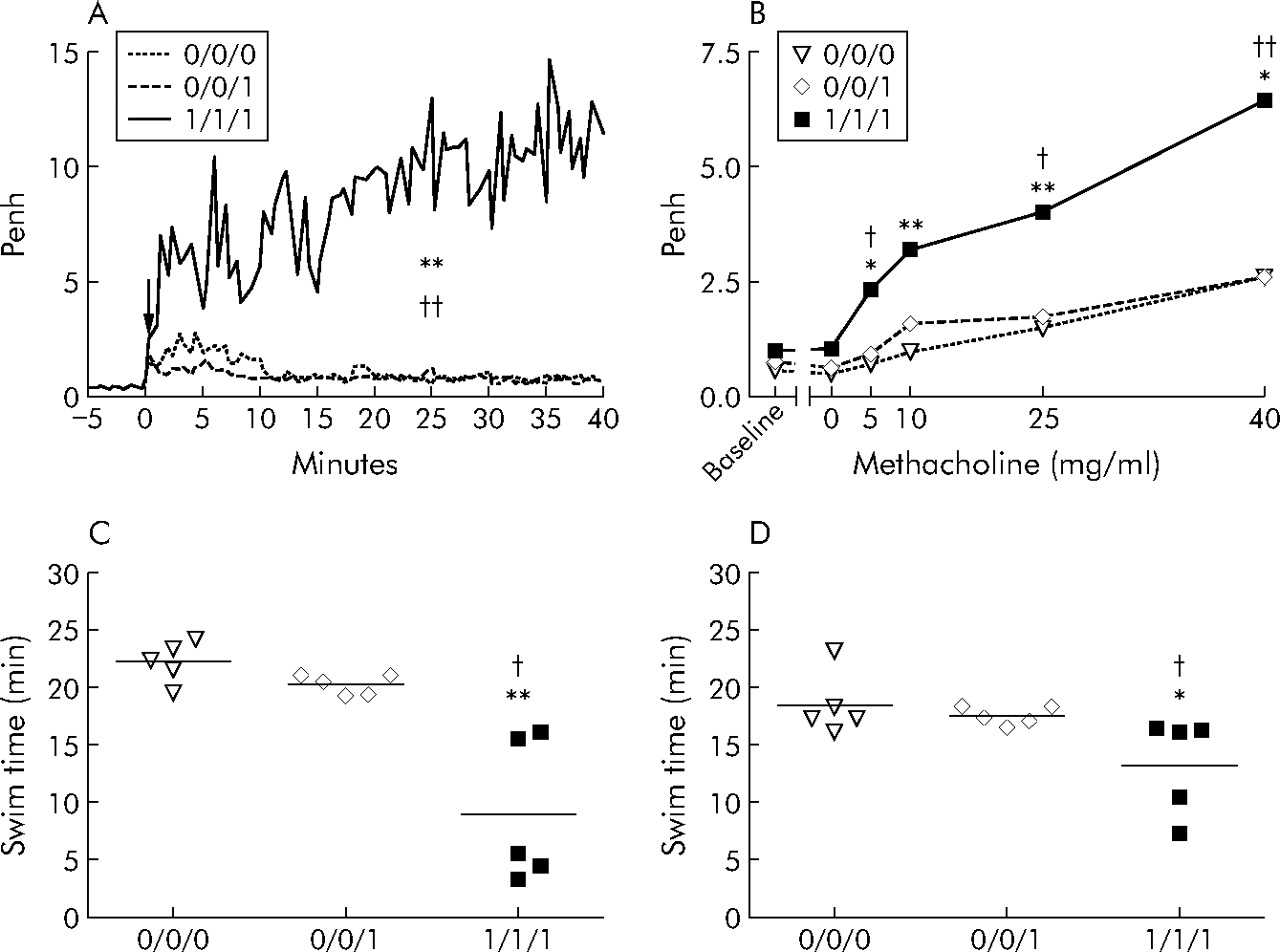
Figure 1⇓ shows the early increase in Penh (panel A) and late increase in methacholine reactivity (panel B) in mice sensitised and challenged with TDI (group 1/1/1) compared with non-sensitised mice that received TDI (0/0/1) and control mice (0/0/0). These changes are similar to those of our previous experiments.2,3 The outcomes of the swimming test parallel those of ventilatory function: in the 1/1/1 group endurance was reduced by 13 minutes (panel C) and by 5 minutes (panel D) at the early and late time points, respectively, compared with the 0/0/1 group whose swimming times did not differ from those of the 0/0/0 group. Additionally, the swim time of the control mice was comparable to those of Matsumoto et al.6 When looking at responses in individual mice, the magnitude of the impairment in the first swim test correlated (non-parametric Spearman correlations) well with the magnitude of the early increase in Penh (r = −0.84, p<0.001) and the late increase in methacholine reactivity (r = −0.72, p<0.01). The second swim test correlated also with the early increase in Penh (r = −0.85, p<0.001) and the increased methacholine reactivity (r = −0.83, p<0.001).
(A) Ventilatory response (Penh) before and after intranasal instillation (arrow) with TDI or vehicle. (B) Methacholine responsiveness (Penh) 24 hours after instillation of TDI or vehicle. (C) Swim test 1 hour after instillation with TDI or vehicle. (D) Swim test 22 hours after instillation with TDI or vehicle. Experimental groups are identified by three symbols. 0 and 1 represent administration of vehicle (acetone:olive oil) or TDI, respectively; the first two symbols identify the agent applied dermally on days 1 and 8, and the third symbol identifies the agent instilled intranasally on day 15. n = 5 per group. †p<0.05, ††p<0.01 compared with the 0/0/1 control group; *p<0.05, **p<0.01 compared with the 0/0/0 control group (non-parametric Kruskal-Wallis test, Dunn’s multiple comparison post hoc test).
This is the first study to use an exercise test to measure the physiological response in a mouse model of asthma. In mice that had been dermally sensitised and then intranasally challenged with TDI, there was an early decrease in endurance 1 hour after challenge and this was still present 1 day later, though to a lesser degree. As in our previous experiments,3 the physiological response depended on the prior dermal administration of TDI, thus excluding a non-specific toxicity of TDI. Although our findings provide further evidence of a relevant functional response in our asthma model, it remains to be verified whether the changes in Penh and in exercise capacity found here are due to reduced nasal patency, bronchoconstriction, or other effects in small airways.
Acknowledgments
The authors thank Professor T Troosters for his comments on the manuscript.