Thorax

HOME HELP FEEDBACK SUBSCRIPTIONS ARCHIVE SEARCH TABLE OF CONTENTS REGISTER
[Advanced]

This Article
Right arrow Extract Freely available
Right arrow Full Text (PDF)
Right arrow Alert me when this article is cited
Right arrow Alert me if a correction is posted
Services
Right arrow Email this link to a friend
Right arrow Similar articles in this journal
Right arrow Similar articles in PubMed
Right arrow Add article to my folders
Right arrow Download to citation manager
Right arrowRequest Permissions
Citing Articles
Right arrow Citing Articles via Google Scholar
Google Scholar
Right arrow Articles by Annema, J T
Right arrow Articles by Rabe, K F
Right arrow Search for Related Content
PubMed
Right arrow PubMed Citation
Right arrow Articles by Annema, J T
Right arrow Articles by Rabe, K F
Topic Collections
Right arrow Thorax Images in Thorax
Thorax 2004;59:636
© 2004 BMJ Publishing Group Ltd & British Thoracic Society


Images in Thorax

A rare cause of Ortner’s syndrome (cardiovocal hoarseness)

J T Annema1, J J Brahim2, K F Rabe3

1 Leiden University Medical Center, Leiden, The Netherlands
2 Leyenburg Hospital, The Hague, The Netherlands
3 Leiden University Medical Center, Leiden, The Netherlands

Correspondence to:
Correspondence to:
Dr J Annema
Department of Pulmonology, P O Box 9600, University Medical Centre, 2300 RC Leiden, The Netherlands; j.t.annema{at}lumc.nl

A 78 year old man with a history of hypertension, arteriosclerosis, and a myocardial infarction woke up one morning to realise that he had lost his voice. He did not feel ill nor had he experienced symptoms of cough or fever. He had smoked cigarettes for over 60 years and had a brother with lung cancer. Left vocal cord paralysis was assessed at laryngoscopy.

A contrast enhanced computed tomographic (CT) scan of the chest showed a mass with a central opacity in the aortopulmonary window (fig 1Go, arrow). In the diagnostic workup densities of this mass were measured which did not correspond with contrast as seen in the aorta. A mediastinal metastasis or mediastinal tumour was the most likely diagnosis given by the radiologist. As he was not aware of the clinical entity of Ortner’s syndrome, an aneurysm was considered less likely. Transoesophageal endoscopic ultrasonography revealed a saccular aneurysm of the aortic arch surrounded by a haematoma (fig 2Go). Using colour Doppler, flow was detected through a 5 mm connection between the aorta and the aneurysm (fig 2Go, arrow).


View larger version (124K):
[in this window]
[in a new window]
 
Figure 1  
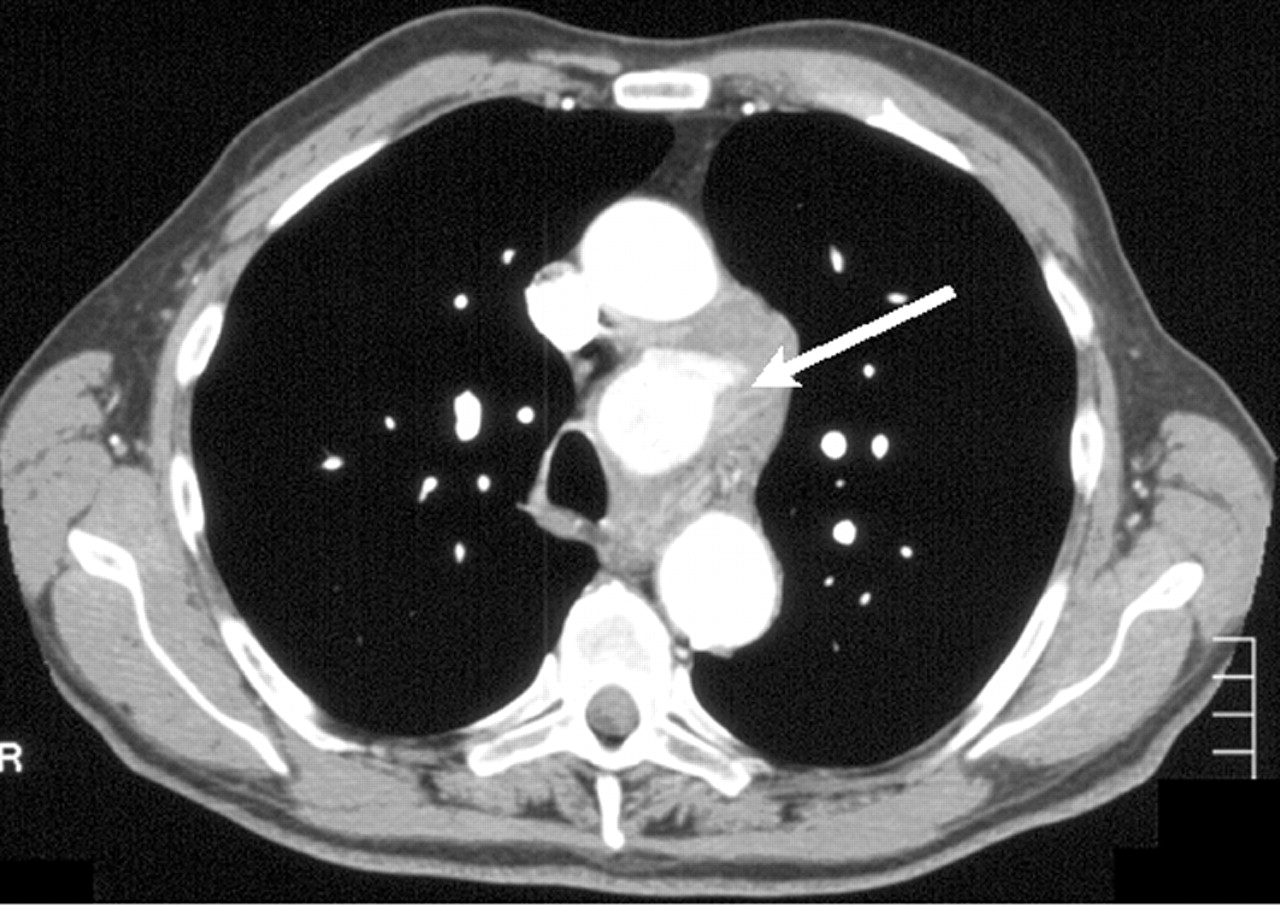
Transverse CT scan of the chest showing a mass (arrow) in the aortopulmonary window.


View larger version (152K):
[in this window]
[in a new window]
 
Figure 2  Transoesophageal echo image showing a saccular aneurysm (An) of the aortic arch surrounded by a haematoma. The arrow points to the connection between the aorta (Ao) and the aneurysm. Es = oesophagus.

 
Only a few such cases have been described, possibly not as well documented as this one. An aortic arch aneurysm is a rare cause of cardiovocal hoarseness, also known as Ortner’s syndrome, first described in 1897 in patients with left atrial enlargement due to mitral valve stenosis.1 In this syndrome the left recurrent laryngeal nerve is thought to be injured due to compression caused by changes in the anatomy of the heart or great vessels, most often by left atrial enlargement due to mitral valve stenosis. Endoscopic ultrasonography may be the diagnostic procedure of choice in establishing the diagnosis.


REFERENCE

  1. Ortner NI. Recurrenslähmung bei Mitralstenose. Wien Klin Wochenschr 1897;10:753–5.


This Article
Right arrow Extract Freely available
Right arrow Full Text (PDF)
Right arrow Alert me when this article is cited
Right arrow Alert me if a correction is posted
Services
Right arrow Email this link to a friend
Right arrow Similar articles in this journal
Right arrow Similar articles in PubMed
Right arrow Add article to my folders
Right arrow Download to citation manager
Right arrowRequest Permissions
Citing Articles
Right arrow Citing Articles via Google Scholar
Google Scholar
Right arrow Articles by Annema, J T
Right arrow Articles by Rabe, K F
Right arrow Search for Related Content
PubMed
Right arrow PubMed Citation
Right arrow Articles by Annema, J T
Right arrow Articles by Rabe, K F
Topic Collections
Right arrow Thorax Images in Thorax


HOME HELP FEEDBACK SUBSCRIPTIONS ARCHIVE SEARCH TABLE OF CONTENTS REGISTER
Terms and conditions relating to subscriptions purchased online  ¦  Website terms and conditions  ¦  Privacy policy
Copyright © 2004 BMJ Publishing Group Ltd & British Thoracic Society